You walk out of the doctor's surgery clutching a medical report bearing the word "dermatitis". Moments later, the pharmacist hands you a soothing ointment for your "eczema". Meanwhile, all you can see in the mirror is a red, angry, intensely itchy rash. Three different words for what looks like the exact same problem—no wonder patients feel bewildered. This terminology trap is a real source of frustration, especially when you find yourself asking, "what type of eczema do I have?" and your current treatments simply aren't working. Exploring the different types of eczema and understanding their characteristics is the first step towards finding genuine relief. While most online guides cover only the seven most common variations, modern medicine actually recognises a much broader spectrum of skin inflammations. These conditions can look remarkably similar to the untrained eye, but they stem from entirely different root causes. If you apply a treatment meant for an allergic reaction to a yeast-driven problem, your skin won't improve. Your body isn't working against you; it's simply reacting to a specific internal or external trigger. This guide gives you an accessible yet medically precise overview of all 12 types of eczema. By the end of this article, you'll finally understand the language your skin is speaking.
Key takeaways if you're short on time
- Accurate diagnosis dictates treatment success, because the wrong ointment can actively worsen your condition; this is why correctly identifying the types of eczema matters so much.
- Eczema and dermatitis are essentially the same thing, as both terms are used interchangeably in clinical practice to describe skin inflammation, with specific types set apart by adjectives (such as atopic, contact, or seborrhoeic).
- The root cause lies both inside and outside the body, with inflammation usually arising from a combination of a compromised skin barrier, specific immune system reactions, and microbiome imbalances.
- Itching follows different biological pathways, which means that while antihistamines work wonders for hives, they do nothing for the non-histamine itch of neurodermatitis or atopic eczema.
- Good basic skincare unites all diagnoses, as restoring the lipid layer and washing gently will help soothe the skin whatever your exact medical condition.
Eczema vs Dermatitis: Why They Are (Almost) the Exact Same Thing
One of the most frequent questions patients ask is whether they are dealing with eczema or dermatitis. In clinical dermatology, the two terms are functionally synonymous. The word "dermatitis" comes from Greek and translates literally to inflammation of the skin (derma = skin, itis = inflammation). The term "eczema" comes from the Greek word "ekzein", meaning "to boil over" or "to erupt". If you have ever had an acute flare-up complete with tiny, fluid-filled blisters, you'll know how accurate that ancient description is.
Doctors generally divide these inflammatory conditions into two broad categories. Exogenous types of eczema are triggered by the external environment. This category includes contact reactions to certain metals, harsh chemicals, or chronic irritation from constant exposure to water. Endogenous types of eczema, on the other hand, are driven by internal factors—typically a genetic predisposition or a specific dysregulation of the immune system. Atopic and seborrhoeic eczema fall firmly into this latter group.
The physical symptoms shift dramatically depending on the stage of the condition. Acute eczema is marked by weeping, oozing, intense redness, and the formation of tiny vesicles (blisters). Chronic eczema, by contrast, dries out. The skin becomes rough, thickened, and scaly, developing deep fissures—a process dermatologists call lichenification, which essentially means the skin thickens so much that its natural lines become deeply exaggerated.
On a cellular level, eczema rests on three main pillars. The first is a badly compromised skin barrier that lacks essential ceramides (the lipid mortar that holds skin cells together) and structural proteins such as filaggrin. The second is immune dysregulation, where white blood cells overreact to perfectly normal, everyday stimuli. The third pillar is a disrupted microbiome, which lets pathogenic bacteria such as Staphylococcus aureus (golden staph) colonise and dominate the skin's surface. You can explore the mechanisms behind these skin reactions in our detailed guide on What causes an itchy rash.
Overview Table of the 12 Types of Eczema
| Type | Primary characteristic | Typical location | Prevalence |
|---|---|---|---|
| Atopic eczema | Itching + dry skin + thickening (lichenification) | Creases of elbows and knees, face | ~11% of children, ~6% of adults globally (Dermatitis 2024) |
| Contact eczema (ICD+ACD) | Redness and blisters after contact | Hands, face, neck | 20.1% (WAO 2025) |
| Seborrhoeic eczema | Greasy scales, yellowish crusts | Scalp, face, chest | 1–5% (135.7 million GBD 2021) |
| Dyshidrotic eczema | Tiny "tapioca-like" blisters | Palms, fingers, soles of feet | 5–20% of hand eczemas |
| Nummular eczema | Coin-shaped lesions | Lower legs, torso | 0.07–0.26% (JDDG 2025) |
| Varicose (stasis) eczema | Brown pigmentation, swelling | Lower legs | 5.9–6.9% in over-50s |
| Asteatotic eczema | "Cracked porcelain" appearance | Lower legs, arms | > 40% of elderly in care (Japan) |
| Neurodermatitis (LSC) | Thickened patches from chronic scratching | Neck, ankles, genitals | ~12% |
| Perioral dermatitis | Tiny spots and pustules, spares the lip border | Around the mouth and nose | 90% are women aged 16–45 |
| Hives (acute urticaria) | Raised wheals with pale centres | Whole body | 20% lifetime risk |
| Chronic urticaria (CSU) | Wheals lasting > 6 weeks | Whole body | 1.4% |
| Occupational eczema | Work-related skin damage | Hands, forearms | Up to 38% in high-risk professions |
This table serves as a quick reference for initial orientation. The following sections break down each of the different types of eczema in detail, including the latest scientific advances in their treatment.
Contact Eczema: When Your Body Says "Stop"
Contact eczema splits into two completely different biological processes: allergic contact dermatitis (ACD) and irritant contact dermatitis (ICD). Telling them apart determines whether your treatment will succeed or fail.
Allergic contact eczema relies on immunological memory. It is classified as a delayed-type hypersensitivity reaction. Unlike a pollen allergy that triggers sneezing within minutes, this skin reaction takes 48 to 72 hours to appear after you touch the offending substance. Tiny molecules known as haptens penetrate the skin, bind to your natural proteins, and alert your immune cells, which then memorise this specific structure. The next time you encounter that exact allergen, your immune system launches a powerful, destructive inflammatory attack. Irritant eczema works differently. It is caused by the direct toxic effect of harsh chemicals—such as solvents or strong surfactants (like SLS, sodium lauryl sulphate, found in many foaming cleansers)—that physically strip away and dissolve the skin's protective lipid barrier. It needs no prior sensitisation and will affect anyone exposed to the irritant for long enough.
In Europe, the most common allergen remains nickel, which sensitises 18.1% of the population. Fragrances follow closely at 16% (with oxidised linalool alone accounting for 11.1%). Reassuringly, the infamous preservative methylisothiazolinone has dropped from 8% to 2.9% following a strict EU ban in 2017. A rising concern in 2024 is the surge in allergies to CEDMC, a chemical accelerator used to make nitrile gloves.
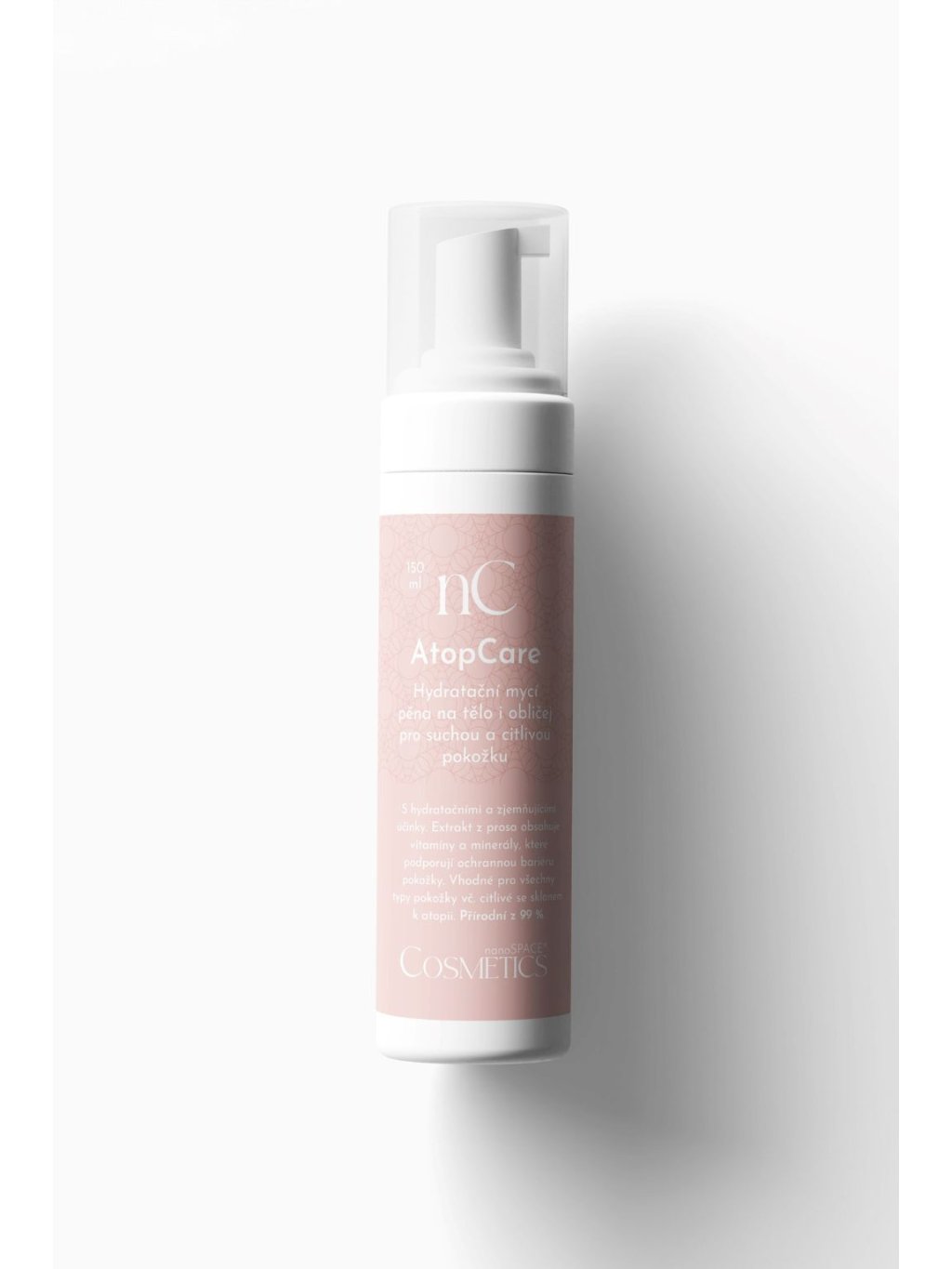
Diagnosis is carried out by a dermatologist using patch testing. The standard European baseline series (ESCD) tests for the 32 most prevalent allergens. Treatment demands complete elimination of the trigger, bearing in mind that a badly damaged skin barrier can take up to six months to fully repair. During the acute weeping phase, doctors prescribe topical corticosteroids, reserving systemic drugs such as alitretinoin for severe, unmanageable cases. To support barrier repair during this long healing window, an intensive barrier cream such as the AtopCare hand cream is invaluable.
Modern medicine has recently delivered a real breakthrough in the form of JAK inhibitors. In 2025, the US FDA approved the drug delgocitinib (marketed as Anzupgo) as the first targeted therapy specifically for chronic hand eczema. The most severe, life-altering forms are also treated with the biologic drug dupilumab.
See a doctor straight away if the reaction spreads beyond the original area of contact, if blisters erupt on your face, or if the rash fails to respond to basic care within two weeks. To understand how this relates to broader systemic reactions, read our guide on an Itchy rash all over the body.
Seborrhoeic Eczema: Dandruff as a Symptom, Not a Diagnosis
According to data from the Global Burden of Disease study (GBD 2021), seborrhoeic eczema affects 135.7 million people worldwide, a 53% increase since 1990. Countless patients wrongly believe that the characteristic dandruff and facial redness are the result of poor hygiene. The reality is quite the opposite.
The pathophysiology of seborrhoea lies in a very specific, dysfunctional interaction between the skin and its microbiome. Yeasts of the genus Malassezia live naturally on everyone's skin. In healthy people, they do no harm. In patients with seborrhoea, however, these yeasts break down the skin's natural sebum (oil), producing irritating oleic acid as a byproduct. This acid penetrates the top layers of the skin and triggers an inflammatory cascade. The skin responds by sharply accelerating its cell turnover, which leads to the rapid build-up of greasy, yellowish scales.
Typical locations are areas with the highest density of sebaceous (oil) glands. These lesions commonly appear on the scalp, in the nasolabial folds (smile lines), in the eyebrows, and on the centre of the chest. Interestingly, on darker skin tones the classic red erythema is often absent, and the lesions show a purplish or violaceous discolouration instead. Gentle, non-stripping cleansing is crucial here; using a product such as the AtopCare cleansing foam helps maintain hygiene without provoking further oil production.
Medical research has also uncovered a fascinating neurological connection. Seborrhoeic dermatitis occurs in 18.6–59% of patients with Parkinson's disease, compared with just 1–5% in the general population. It is now being studied as a possible early biomarker for this neurological condition.
Standard treatment involves topical antifungals, typically 2% ketoconazole or ciclopirox. The year 2023 brought a major step forward with the approval of a 0.3% roflumilast foam. In clinical trials, 80% of patients achieved an excellent score, and 50% had completely clear skin within eight weeks. Seek medical advice if the rash spreads, if weeping erosions appear, or if over-the-counter shampoos fail to bring relief within four weeks. For a deeper dive into managing this condition, explore our article: What is seborrhoeic dermatitis.
Dyshidrotic Eczema: Tiny Blisters, Immense Frustration
The very name of this condition is rooted in a historical medical error. The word "dyshidrotic" implies a disorder of sweating. Today, science has conclusively shown that sweat glands have nothing to do with the formation of these very specific blisters. Understanding this one fact helps patients stop blaming themselves for having sweaty hands and allows them to adopt the right care strategy.
This distinct type accounts for 5–20% of all hand eczemas. It presents as a sudden outbreak of hard, deep-seated vesicles measuring 1–2 millimetres across. They closely resemble tapioca pearls and typically erupt on the lateral (side) aspects of the fingers, the palms, and the soles of the feet. The itching is often described as excruciating. These blisters do not pop easily; instead, they slowly dry out over the course of two to three weeks, leaving behind very dry, peeling, fissured skin.
The triggers are notoriously elusive. A frequent hidden culprit is a contact allergy to metals. Nickel and cobalt often work in tandem—up to 25% of patients allergic to nickel also react to cobalt. Another surprising trigger is a seemingly unrelated fungal infection on the feet (tinea pedis). The body generates what is known as an "Id reaction", essentially a remote allergic response. Curing the athlete's foot clears the hand eczema in a full third of all dyshidrotic cases.
Dermatologists typically reach for ultra-potent topical corticosteroids, as the skin on the palms is exceptionally thick and hard to penetrate. For severe, stubborn cases, PUVA phototherapy or botulinum toxin injections prove highly effective (since up to seven out of 10 patients also suffer from hyperhidrosis). The systemic drug alitretinoin shows excellent results, though efficacy drops to 44.4% in patients of Asian descent. 2025 brings delgocitinib (Anzupgo) to the market, the first FDA-approved medication specifically for chronic hand eczema, which showed remarkable efficacy in the DELTA 1 and 2 trials published in The Lancet. See a doctor if your eczema relapses more than three times a year or if deep, painful cracks develop. Learn more in our dedicated guide: Dyshidrotic eczema: causes and symptoms.
Nummular Eczema: The Coin-Shaped Lesions That Mimic Atopy
Nummular eczema (also known as discoid eczema) is marked by sharply defined, coin-shaped lesions that itch maddeningly. It mainly affects men aged between 50 and 65. The distinct circular patches appear chiefly on the lower legs and torso, and they have a strong tendency to weep, ooze, and form thick crusts.
In 2023, an important reclassification of this disease took place. A landmark study published in the Journal of Allergy and Clinical Immunology (Bohner et al.) officially designated nummular eczema as the "neglected twin" of atopic dermatitis. Both diseases share a marked dysfunction of the skin barrier, eosinophilia, and heavy colonisation by golden staph bacteria. The true difference only reveals itself under a microscope. The histological picture of nummular eczema actually looks closer to psoriasis, as it shows a co-dominant inflammation driven by both the Th2 and Th17 immune pathways.
The primary trigger is very dry skin, which plagues two-thirds of all patients. A cold climate, frequent bathing in scalding hot water, and local mechanical trauma (such as a minor graze or an insect bite) can instantly provoke a brand-new coin-shaped lesion. Regular, generous moisturising with a rich formula such as the AtopCare nourishing body cream is vital for prevention.
Treatment demands potent topical corticosteroids because these lesions are notoriously stubborn and resistant. Research into systemic drugs has produced intriguing data. While apremilast proved ineffective (a 2025 TU München study showed improvement in only 6.7% of patients compared with 25% on placebo), biologic therapy with dupilumab has brought lasting skin clearance in recent case studies. A doctor's visit is essential if the lesions start to spread quickly, primarily to distinguish the condition from a fungal ringworm infection (tinea corporis), which looks identical to the untrained eye.
Varicose Eczema: A Venous Cause, a Skin Symptom
Also known as stasis dermatitis, this type of eczema affects 5.9–6.9% of the population over the age of 50, with prevalence climbing past 20% in the over-70s. Crucially, this is not primarily a skin disease; it is the direct consequence of a failing vascular system.
The pathophysiology begins with chronic venous insufficiency. The tiny, one-way valves inside the veins of the lower legs fail to close properly. Blood pools and stagnates, which raises the pressure inside the veins (from a normal resting pressure of under 30 mmHg to as much as 80–90 mmHg). This extreme hydrostatic pressure literally forces white blood cells out of the blood vessels and into the surrounding tissue, where they become trapped and fuel chronic inflammation. At the same time, red blood cells break down and deposit an iron-rich pigment called haemosiderin into the skin. This causes the classic, rust-brown hyperpigmentation that doctors look for. The inflammatory distress signals in the skin then trigger relentless itching.
Left untreated, the condition spirals into serious complications. The subcutaneous fat hardens and scars (a process called lipodermatosclerosis). The skin, starved of oxygen and nutrients, eventually breaks down into open venous leg ulcers, which account for 60–80% of all lower-limb ulcers and carry an 80% risk of recurrence within three months. Up to 37% of patients also suffer from autoeczematisation, where the rash suddenly spreads across the entire body.
Clinical practice highlights one major, hidden danger. Up to 46.7% of patients develop a contact allergy to the very medicinal ointments prescribed to heal them. They frequently react to bacitracin, balsam of Peru, or standard preservatives. The choice of emollients must therefore follow strict rules; pure, preservative-free pharmacy-grade petrolatum is often the safest option. The cornerstone of treatment remains compression therapy (bandages or stockings), even though patient compliance stubbornly hovers at a mere 50–60%. See a doctor straight away if an open ulcer forms or if you notice progressive hardening of the skin around your ankles.
Asteatotic Eczema: Eczema From Drought, Not Allergy
Asteatotic eczema is the single most common eczematous condition among the elderly, with a median onset age of 69. In Japanese long-term care (LTC) facilities, prevalence reaches 41.2%. It is not caused by an allergy, nor is it an autoimmune condition; it is caused by extreme skin dehydration.
The clinical picture is distinctive and hard to mistake. The skin resembles cracked porcelain or the parched bed of a dried-up lake—a presentation that dermatologists officially term "crazy paving". The lower legs and arms become covered in a network of fine, red fissures that divide the skin into tiny, dry, diamond-shaped plates.
As we age, our natural production of epidermal lipids (ceramides, cholesterol, and free fatty acids) plummets. At the same time, the cells lose their ability to bind and hold on to water—they simply let it evaporate. Transepidermal water loss (TEWL) can spike up to 75 times above normal, healthy levels. The uppermost layer of the skin begins to shrink, contract, and mechanically crack open. These microscopic fissures act as open doors, letting everyday environmental irritants penetrate deep into the dermis, instantly triggering an inflammatory response.
Dermatologists warn of one rare but very dangerous phenomenon. If asteatotic eczema suddenly erupts and spreads rapidly across the entire body of an elderly patient and fails to respond to strong corticosteroids, doctors must urgently rule out T-cell lymphoma or Hodgkin's lymphoma. In rare cases, this sudden, severe dryness can be a cutaneous sign of a hidden internal malignancy (known as a paraneoplastic syndrome).
Standard treatment calls for aggressive, heavy-duty lipid replenishment. Applying a rich emollient such as the AtopCare body oil to damp skin within three minutes of stepping out of the shower physically locks the water inside the epidermis. Modern therapeutic approaches use creams containing N-palmitoylethanolamine to soothe the nerves. The gold standard in Japan involves heparinoid creams (Hirudoid 0.3%), which Japanese literature shows induce remission in up to 95% of patients. See a doctor if deep fissures appear on the face or if the condition spreads across the body. To help you choose the right lipid care, read our guide on What actually works for extremely dry skin.
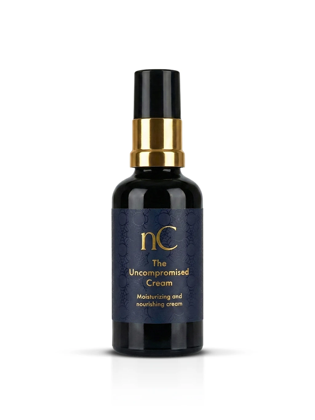
Editor's pick
The Uncompromised Cream
An exceptional anti-ageing day cream from nanoSPACE Cosmetics, expertly formulated for mature, dry, or highly sensitive skin that needs intensive nourishment with no compromises on its ingredients.
€56
View productNeurodermatitis: The Vicious Cycle of Itching and Scratching
Medical terminology adds yet another layer of confusion here. In German-speaking countries, the term "Neurodermitis" is widely used by the public to mean atopic eczema. In British and international medical literature, however, neurodermatitis refers to something entirely different—a chronic condition officially known as Lichen Simplex Chronicus (LSC).
The prevalence of LSC reaches roughly 12% of the population. It is a highly localised dermatosis with one very specific diagnostic hallmark. The thickened, leathery plaques form exclusively in areas the patient can physically reach with their hands—typically the nape of the neck, the ankles, the forearms, or the genitals. You'll never find an LSC patch in the middle of the back, between the shoulder blades. The reason lies entirely in how the disease develops.
The condition is driven by an unbreakable vicious cycle. The patient feels a mild itch and scratches it, microscopically damaging the top layer of the skin. The damaged skin releases inflammatory cytokines, which activate local nerve endings and signal the brain to produce even more itching. Over time, the brain memorises this repeated stimulation and becomes hyper-reactive—a phenomenon neurologists call central sensitisation. The physical act of scratching brings strong, if only temporary, psychological relief, which patients rate as 3.4 out of 5 on clinical reward scales. This powerful behavioural mechanism can keep the local inflammation burning for years. Here's a vital piece of information for frustrated patients: this specific itch is not caused by histamine. Standard over-the-counter antihistamines are therefore useless, and taking them will bring you no relief at all.
The psychological burden is heavy. Patients with clinical anxiety have a 41-fold higher risk of developing LSC, and 62% of diagnosed patients have a concurrent psychiatric diagnosis. Treatment requires forcefully breaking the cycle. Dermatologists often apply a potent corticosteroid under a strict occlusive dressing (such as a bandage) to physically stop the patient's fingernails from reaching the skin. To dampen the neuropathic itch signals, doctors may prescribe nerve-calming medications such as gabapentin or duloxetine, ideally combined with cognitive behavioural therapy (CBT) to unlearn the scratching habit. At present, there are no FDA-approved drugs designed specifically for LSC. See a doctor if diligent moisturising and sheer willpower cannot break the itch-scratch cycle within two weeks. We explore these psychological triggers further in our article: Understanding the stress rash.
Perioral Dermatitis: The Paradox Caused by the Very Cream Meant to Heal It
Up to 90% of those affected by this condition are women aged between 16 and 45. Perioral (and sometimes periorbital) dermatitis appears as a sudden breakout of tiny red papules and pus-filled pustules clustered around the mouth, nose, or eyes. A careful clinical examination reveals a telling diagnostic detail that distinguishes the disease from adult acne. The rash always strictly spares the vermilion border—leaving a narrow, pale, completely healthy ring of skin directly next to the lips.
The primary cause of this condition is a cruel dermatological paradox. The disease is most often triggered by the inappropriate use of topical corticosteroids on the delicate skin of the face. The biological mechanism is exceptionally well documented. The steroid cream inhibits the production of nitric oxide (NO) in the skin, which forces the local blood vessels to constrict tightly (vasoconstriction). For a time, the skin looks pale, clear, and seemingly cured. The moment the patient stops applying the cream, however, a sharp rebound dilation occurs. The blood vessels open wide, blood floods the tissue, and angry inflammatory pustules erupt. Alarmed by the sudden breakout, the patient immediately reaches for the steroid cream again. This creates a genuine physical addiction within the skin tissue. The skin's microbiome also plays a defining role, as the disrupted barrier allows Fusobacterium and Demodex skin mites to multiply rapidly.
Treatment relies on a demanding process known as "zero therapy". This means the radical, cold-turkey withdrawal of all cosmetics, moisturisers, and, above all, all steroid creams. The patient must be warned firmly in advance: the skin will look and feel noticeably worse during the first few days of withdrawal. To help bridge this difficult acute phase, doctors prescribe topical metronidazole 0.75%, 20% azelaic acid, or pimecrolimus cream. Severe cases call for a course of oral antibiotics from the tetracycline family, lasting eight to 12 weeks. The year 2025 brings new hope with a 0.3% roflumilast cream, which in recent trials cleared the skin in just five days and maintained remission for 11 months. Don't delay seeing a doctor; this diagnosis must be confirmed by a specialist who can safely guide you through the difficult process of steroid withdrawal.
Hives: An Allergic Reaction or a Chronic Mystery?
Hives, medically known as urticaria, will affect up to 20% of the population in their acute form (lasting less than six weeks) at some point in their lives. The chronic form (CSU), however, affects 1.4% of people, which works out at 65.1 million long-suffering patients worldwide.
The underlying pathophysiological process is the degranulation of mast cells. These specialised immune cells rupture and spill histamine and inflammatory cytokines straight into the surrounding tissue. The immediate result is the formation of wheals—raised, intensely itchy, swollen patches with a pale centre and a bright red halo. These wheals are notoriously transient; they travel across the body, change shape, and vanish without leaving a single scar within 24 hours.
Chronic spontaneous urticaria (CSU) divides into two distinct subtypes. Type I (auto-allergic) accounts for 70–80% of cases, and these patients respond beautifully to targeted biologic therapy with omalizumab. Type IIb, however, has a darker autoimmune foundation—the body mistakenly creates antibodies that attack its own mast cells. This type has a much more severe clinical course, responds very poorly to standard antihistamines, and often requires powerful immunosuppressants such as ciclosporin.
Up to a third of patients with CSU also experience angioedema, a deep, dangerous swelling of the subcutaneous tissue. A doctor must quickly distinguish between two types of angioedema. Histaminergic angioedema responds to antihistamines and typically swells the face, lips, and tongue. Bradykinin-mediated angioedema, on the other hand, is frequently triggered by common blood pressure medications (ACE inhibitors) and needs an entirely different, life-saving emergency treatment. A very specific subgroup is chronic inducible urticaria (CIndU), which is triggered by physical stimuli. Cold urticaria, for instance, carries a serious, life-threatening risk of anaphylactic shock if the patient swims in cold water. Call for emergency medical help immediately if you experience any swelling of the face or airways. Chronic forms are managed by a dermatologist or immunologist. We explain these mechanisms in greater detail in our guide on Allergic rashes and hives.
Occupational Eczema: The Heavy Price of "Wet Work"
Working with your hands takes a heavy toll. Occupational eczema plagues very specific industries. Among healthcare workers, patch test positivity reaches a worrying 31.5%. Hairdressers face a lifetime prevalence of hand dermatitis of 38.2%. For construction workers, the incidence hits 36.9%, with the primary culprit being hexavalent chromium found in wet cement, which is also a potent carcinogen.
Dermatology defines the concept of "wet work" with strict, measurable parameters: any occupation where a worker's hands are immersed in a wet environment for more than two hours per day, or where they are forced to wash their hands more than 20 times per shift. This constant soaking and drying physically destroys the lipid barrier, turning the skin into a sponge that absorbs industrial allergens straight into the deep dermal layers.
In occupational settings, prevention matters far more than treatment. The foundation is applying heavy barrier creams before the shift begins, wearing breathable cotton liners underneath protective nitrile or latex gloves, and strictly cutting out any unnecessary hand washing. Occupational eczema is not merely a medical issue; it's a complex legal one. It is an officially recognised occupational disease that can ultimately force a patient to give up their chosen profession altogether. You should consult an occupational health doctor if the condition of your skin stops you from doing your job safely. For strategies on healing a destroyed barrier, read our article on Cracked skin on hands, or explore our broader guide on hand eczema.
Atopic Eczema: The Largest Member of the Family
Atopic eczema (atopic dermatitis) is a chronic, highly inflammatory skin disease with a strong genetic foundation. The central player in this condition is a genetic mutation affecting the production of filaggrin (FLG), a vital structural protein that binds skin cells together to form a watertight seal. Without enough filaggrin, the skin simply cannot hold on to moisture; it dries out quickly and leaves the gates wide open for external allergens and bacteria to invade.
Crucially, this disease does not exist in isolation. It is inextricably linked to a cascade of comorbidities known as the "atopic march". Children with severe atopic eczema have a statistically high probability of going on to develop food allergies, allergic rhinitis (hay fever), and bronchial asthma as they grow older. The clinical course is notoriously exhausting, marked by a relentless cycle of quiet periods (remission) shattered by sudden, violent flare-ups (exacerbations).
Given the biological complexity and emotional weight of this disease, we've dedicated an entire series of in-depth articles specifically to atopic eczema. For concrete, actionable strategies on how to suppress the maddening itch and prolong your periods of remission, read our comprehensive guide on Effective treatments for atopic eczema. We also carefully break down the unique challenges of paediatric skin and the emotional hurdles of caring for the youngest patients in our dedicated article on Atopic eczema in children. Finding the right lipid care is vital, and a targeted emollient routine can change your life.
Skincare for eczema-prone skin — the AtopCare range


Common Care Principles: 4 Essential Rules for All Types of Eczema
Although the pathophysiology of each individual disease differs dramatically, the foundation of dermatological care rests on a few unshakeable pillars. Master these four biological principles and you'll gain real control over the state of your skin, effectively supporting a damaged skin barrier whatever your exact medical diagnosis.
- Rebuild the lipid barrier: Inflammation tears apart the intercellular mortar of your skin. Supplying ceramides and occlusive agents (such as pure petrolatum) artificially replaces these missing skin fats, halts heavy water loss, and physically blocks aggressive bacteria from entering the tissue.
- Get the timing of hydration right: Smearing moisturiser onto bone-dry skin is biologically inefficient. Apply your cream or oil to damp skin, ideally within three minutes of stepping out of the shower. This traps and locks the water deep inside the epidermis.
- Hunt down the trigger: Inflammation doesn't appear out of thin air. Ask your doctor for patch tests to uncover hidden contact allergies, or follow a strict, journal-tracked elimination diet for atopic eczema to identify sneaky food triggers.
- Respect the power of corticosteroids: Steroids are a powerful tool for quickly halting an acute immune reaction, but they demand respect. Never apply them to your face without explicit instructions from a specialist, don't exceed two weeks of continuous application on thin skin, and never use them as a substitute for your daily, heavy-duty moisturising routine.
Conclusion — Eczema Is Not a Diagnosis, It Is a Family of Diagnoses
If you've read this far, you now understand that the terminology confusion between doctors, pharmacists, and the internet is, unfortunately, the normal state of affairs. Dermatology is a constantly evolving field that is still sharpening the boundaries between these complex diseases. "Eczema" is simply not one word for one disease. The different types of eczema form a vast, sprawling family tree of diagnoses, each of which demands a very specific approach, an entirely different medical treatment, and a varying degree of patience.
Armed with the knowledge of your exact type, you hold a real advantage. You can finally stop wasting money on the wrong creams, and you have the clinical vocabulary you need for a genuinely meaningful, two-way conversation with your dermatologist. Find your specific match in the overview above and dive deep. Learn how to conquer the maddening night-time itch of dyshidrotic eczema, discover how to starve the yeast driving your seborrhoeic dermatitis, or finally understand the crucial difference between deep tissue swelling and superficial wheals in our guide to hives. Your skin is trying hard to communicate with you. Now you finally know how to speak its language.
Frequently asked questions
How do I tell the difference between eczema and psoriasis?
Psoriasis creates very thick, silvery-white scales that sit on top of sharply defined, bright red plaques. Crucially, psoriasis never forms weeping, fluid-filled blisters. It typically targets the outer, extensor surfaces of joints (such as the points of your elbows and knees) and the scalp, and it often causes tiny, distinct pits to form on your fingernails. Eczema, by contrast, weeps and oozes clear fluid during its acute phase, and thickens the skin (lichenification) during its chronic phase. It prefers to attack the inner creases of the skin (the crooks of your elbows and the backs of your knees). Sometimes, though, the visual symptoms overlap so closely that a dermatologist must perform a small skin biopsy. This is a standard, safe procedure that gives a precise diagnosis.
When is the right time to finally see a dermatologist?
Seek emergency medical care immediately if you develop angioedema (swelling of the lips, tongue, or face), if pustules erupt across your face, if a rash suddenly spreads over your entire body, or if you suspect a secondary infection (indicated by golden-yellow crusts, skin that is hot to the touch, heavy oozing, and throbbing pain). Book an urgent appointment if your eczema is stopping you from sleeping, limiting your ability to work, or if your skin is cracking painfully. Remember that certain types of eczema (such as varicose, perioral, or asteatotic) call for a radically different medical approach than standard atopic eczema, so an exact diagnosis must always be confirmed by a qualified specialist. If over-the-counter care fails to bring relief within two to four weeks, it's time to escalate.
Do steroid creams cure all types of eczema?
No—different types of eczema react to corticosteroids in very different ways. For asteatotic eczema, a steroid will only briefly suppress the redness but does nothing to fix the severe lack of skin lipids, which is the actual root cause. In the case of perioral dermatitis, the steroid cream is often the very thing that caused the disease in the first place, and continued application leads to a severe physical addiction within the skin. For varicose eczema, steroids must be used with great caution because there is a real risk of causing atrophy (irreversible thinning) of the already fragile, damaged skin on the lower legs. The decision to use a steroid, along with its strength and duration, depends entirely on your precise diagnosis.
Is eczema contagious?
No, it isn't. Not a single type of eczema can be passed on through physical touch, through the air, or by sharing towels or clothing. Even though a severe acute phase—complete with weeping blisters, raw skin, and heavy peeling—might look alarming to others, the person next to you is at no risk of catching it. The only exception to this rule is if the eczema lesions become secondarily infected with a transmissible pathogen, such as the golden staph bacteria or the herpes simplex virus (eczema herpeticum). In those specific cases, however, it's the secondary infection that is contagious, not the underlying eczema itself.
Will my eczema come back after I finish treatment?
This depends entirely on the specific type of disease you have. Allergic contact eczema will never return—provided you successfully identify the exact allergen and manage to avoid it for the rest of your life. Atopic and seborrhoeic eczema, however, are chronic, lifelong conditions; their natural course is defined by alternating periods of complete calm and sudden, frustrating flare-ups. Statistics show that dyshidrotic eczema relapses in 50–60% of all patients. This is exactly why a rigorous daily routine of barrier repair and lipid replenishment is so crucial, as a strong skin barrier can significantly reduce both the frequency and the intensity of any future attacks.

Sources
- Alinaghi, F. et al. (2019) 'Prevalence of contact allergy in the general population: a systematic review and meta-analysis', Contact Dermatitis, 80(2), 77–85.
- GBD 2021 Skin Diseases Collaborators (2025) 'Global epidemiology of seborrhoeic dermatitis 1990–2021', Frontiers in Medicine.
- Bohner, A. et al. (2023) 'Nummular eczema as a phenotype of atopic dermatitis: RNA-sequencing reclassification', Journal of Allergy and Clinical Immunology (JACI).
- Hagenström, K. et al. (2025) 'Prevalence and treatment patterns of nummular eczema in Germany', Journal der Deutschen Dermatologischen Gesellschaft (JDDG).
- Bissonnette, R. et al. (2024) 'Delgocitinib cream for chronic hand eczema (DELTA 1 and DELTA 2)', The Lancet, 404(10455).
- Schäfer, T. et al. (2023) 'Stasis dermatitis and chronic venous insufficiency: pathophysiology and management', Phlebologie.
- Misery, L. et al. (2022) 'Lichen simplex chronicus: pathogenesis and clinical update', Experimental Dermatology, 31(5), 681–689.
- Zuberbier, T. et al. (2022) 'EAACI/GA²LEN/EuroGuiDerm/APAAACI guideline for the definition, classification, diagnosis and management of urticaria', Allergy, 77(3), 734–766.